Ultrananaotech, ULTRANANOTECH
Gold Nanoparticles Conjugate As a Tools For Enhanced Diagnosis And Therapy for Cancer
10
Sep
Sep
MAGNIFYING MEDICINE: Gold Nanoparticle conjugate as tools for Enhanced Diagnosis and Therapy for Cancer
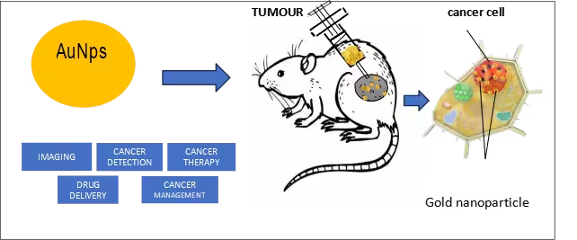
The application of nanotechnology for the treatment of cancer is mostly based on early tumor detection and diagnosis by nanodevices capable of selective targeting and delivery of chemotherapeutic drugs to the specific tumor site. Due to the unique properties of gold nanoparticles like stability, conductivity, biocompatibility etc.., they have been used as a potential tool for the diagnosis of various cancers and drug delivery applications. The non-toxic and non-immunogenic nature of gold nanoparticles and its high permeability in tissues and through tumor cells provide additional benefits by enabling easy penetration and accumulation of drugs at the tumors’ sites. Various innovative approaches with gold nanoparticles are under development. Here, we explore an overview of recent progress made in the application of gold nanoparticles in cancer treatment by tumor detection, drug delivery, imaging and photothermal therapy, as well as their current limitations in bioavailability and applications.
INTRODUCTION
With recent advances in nanotechnology and medical science, numerous nanoparticles and nanomaterials have emerged from different elements such as gold, silver, iron, copper, cobalt, platinum etc., which are synthesized either biologically or physiochemically. Due to low toxicity, large surface-to-volume ratio, and excellent biocompatibility properties of metal nanoparticles, can be used in many possibilities to explore in drug delivery as image contrast agents and for diagnostic purposes.
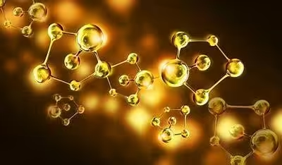
Gold nanoparticles, known for their unique properties like physical, chemical, optical and thermal properties, have opened up a wide range of possibilities in the field of nanoscience and nanotechnology. Among organic and inorganic nanoparticles, gold nanoparticles possess unique optical and surface plasmon resonance properties, it has become the first choice for researchers, mainly in the biology, medicine and pharmaceutical field. These attributes of gold nano with unique optical properties, surface plasmon resonance etc. are utilized in ultrasensitive detection and imaging-based techniques such as cancer. Conventional cancer treatment is based on chemotherapy and radiation, which is treated by using chemotherapeutic drugs or radiation to kill the cancer site through injection of drugs or radiation. This treatment usually causes several side effects due to the damage caused to the surrounding healthy cells
Treating cancer cells, by utilizing a nanoparticle-based drug delivery approach can play a significant role in overcoming the limitations of conventional treatment methodologies and may provide the alternative diagnosis and treatment for cancer.
Numerous medical plants have shown some potential to produce gold nanoparticles within a few seconds, even microorganisms are another powerful source for gold nano particles synthesis, microorganisms are capable of absorbing gold atoms and accumulating the gold nanoparticles by secreting the enzyme and involved in enzymatic reduction of gold ions. Such biologically synthesized gold nanoparticles are used to explore as a tool for biosensors, drug delivery, photoimaging, and photothermal therapy. The biologically synthesized gold nanorods, nanocages, nanotubes, and nanospheres, have become effective tools for human cancer treatment and in various Research and Development of cell biology.
Large surface area and shape selective (triangular) gold nano also plays a significant role in the application in cancer diagnostics, especially in photothermal therapy and photo-imaging. In photo-imaging, triangular shape gold nanoparticles are used because of their higher scattering efficiency, whereas in photothermal therapy, smaller nanoparticles are used because they can absorb light and gold nanoparticles can efficiently converted those absorbed light into heat to destroy the cell
GOLD NANOPARTICLES AS DRUG DELIVERY
Traditional drug delivery approaches for chemotherapeutic drugs through oral or intravenous, which result in the dissemination of the drug in the whole body, with only a fraction of the drug reaching the tumor site. However, this can have side effects on healthy tissues and organs present in the rest of the body.
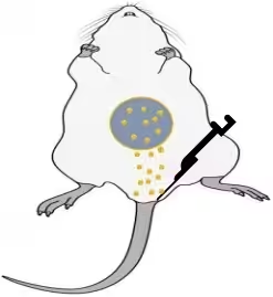
To overcome the limitation of such side effects by targeted drug delivery using nanotechnology approaches, a specific biologically active compound or drug is released at a specific site in a controlled manner. Nanoparticles can efficiently pass through the capillaries to reach the target cells. The chemotherapeutic drugs can be loaded or attached to nanoparticles and can be targeted either passively or actively to the specific site. Tumor tissues generally have a leaky vasculature, which allows the nanoparticles to accumulate easily. This is also known as enhanced permeation and retention
Gold nanoparticles have caught the attention of scientists for their use in drug delivery because of their optical and size tunable properties. They can be prepared in a wide range of core sizes such as 10 to 150 nm, which makes it easier to control their dispersion. Due to the presence of the negative charges on the gold nanoparticles, It can be functionalize easily by the addition of biomolecules e.g. drugs, targeting ligands and genes.
GOLD NANOPARTICLES AS PHOTO IMAGING
Photoimaging is an advanced technique that can help detect early stage of tumors and guide surgeons for precision treatment. Magnetic Resonance Imaging (MRIs) and computed tomography (CT) scans are only used to detect the size of the tumor. Photoimaging is a novel approach in cancer treatment, where millions of functionalized gold nanoparticles are injected into the tumor cell, where they specifically bind to the cancer cells and scatter light, making it easier for the surgeon to identify ttumor cell and healthy cells. Gold nanoparticles such as nanorods, nanocages, and nanoshells are known as the best available photo imaging nanoparticles for cancer therapeutic due to their bio inertness. Some of the applications of photo imaging are:
Surface Plasmon Resonance (SPR) : One of the most important characteristics of the gold nanoparticle is their ability to specific surface plasmon resonance. When gold nanoparticles are exposed to light of specific wavelengths, the collective oscillation of their conduction electrons generates a resonance effect, resulting in enhanced light scattering and absorption. This SPR phenomenon gives rise to strong optical contrast, making gold nanoparticles excellent imaging agents.
Contrast agents: Gold nanoparticles can act as contrast agents in various imaging techniques such as optical microscopy, photoacoustic imaging and medical imaging.
Photoacoustic imaging (PAI): Gold nanoparticles are particularly useful as contrast agents due to their strong ability to convert absorbed light into heat, which generates acoustic waves detectable by ultrasound. This photoacoustic effect enables imaging at deeper tissue depths and makes them valuable for imaging applications in biological tissues.
Two-Photon Imaging: Gold nanoparticles are also used in two-photon imaging, a non linear optical imaging technique that allows imaging at greater depths within tissues. In two-photo imaging, the simultaneous absorption of two low-energy photons excites the nanoparticles, providing better resolution and deeper tissue penetration compared to traditional imaging methods.
Biomedical imaging and nanomedicine: Gold nanoparticles have shown potential in various biomedical imaging applications, such as in vivo imaging of organs, tissues and cells. They are also being investigated for targeting drug delivery in nanomedicine, where imaging capabilities aid in monitoring drug distribution and therapeutic efficacy.
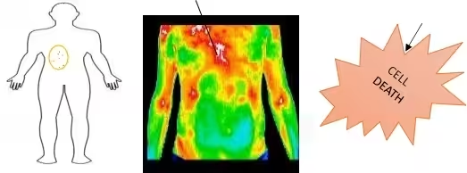
Photothermal Imaging: Gold nanoparticles can be utilized in photothermal imaging, where laser light is used to excite the nanoparticles, causing them to convert light energy into heat. This localized heating effect leads to temperature changes in the sample, which can be detected and used to generate images with high spatial resolution.
GOLD PARTICLES IN BIOMEDICAL APPLICATIONS
IMAGING AND DIAGNOSTIC:
Gold nanoparticles can be functionalized with targeting molecules and imaging agents to mark a specific cell which is affected by tumors and improve detectability and visualization. Gold nanoparticles are used in computed tomography, Fluorescence imaging, and photoacoustic imaging.
DRUG DELIVERY:
Gold nanoparticles serve as a career in drug delivery systems. They can be loaded with therapeutic agents such as drugs, genes, or proteins. Their functionalization is to provide a drug to the specific drug or tissues.
THERAPEUTIC AND TREATMENT:
Gold nanoparticles can exhibit therapeutical properties in certain applications. They are widely used in photothermal therapy, where they absorb light and convert it into energy to selectively destroy the tumor cell
BIOSENSORS AND DIAGNOSTICS:
Gold nanoparticles play a significant role in diagnostic purposes. By using gold with biomolecules, such as antibodies or DNA probes, they can detect specific analytes or biomarkers with high sensitivity and specificity. Gold electrodes are also used in various biosensing devices to measure biomolecular interactions or changes in electrical properties.
TISSUE ENGINEERING AND REGENERATIVE MEDICINE:
The gold nanoparticle can be incorporated into scaffolds or hydrogels used in tissue engineering. Gold nanoparticles have also been investigated for their potential in stimulating tissue regeneration and wound healing processes.
BIOENGINEERING AND BIOCOMPATIBLE MATERIALS:
Gold is compatible with biological systems, making it suitable for fabricating biocompatible materials. It can be used in the product ion of implants, prosthetics, and medical devices due to its corrosion resistance, high biocompatibility, and low reactivity with bodily fluids.
CURRENT LIMITATIONS
COST: Gold is a precious metal, and the production of nanoparticles may be expensive which may limit their widespread use in certain applications.
- STABILITY: In some cases, gold particles can clamp together or aggregate affecting their stability and performance. This aggregation can be influenced by factors such as pH, ionic strength and temperature.
- SIZE DISTRIBUTION: The synthesis of gold nanoparticles can result in the distribution of sizes which can impact their productivity. Achieving a narrow size distribution of crucial for a certain application, especially in drug delivery and imaging.
- TOXICITY: The gold nanoparticles are considered biocompatible their long-term toxicity is still an area of research and concern. The size, shape and surface chemistry of AuNps can influence their interactions with tissues and cells.
- TARGETING EFFICIENCY: As nanoparticles are functionalized with the target delivery, achieving highly specific targets without off-target effects remains a challenge.
- MANUFACTURING SCALABILITY: Scaling up the production of high-quality gold nanoparticles can be challenging and impact their large-scale commercial applications.
- BIOLOGICAL BARRIERS: In vivo applications may face a lot of challenges in biological barriers such as blood-brain barriers, limiting the gold nanoparticles to reach the target tissues or cells.
- REGULAR CONSIDERATIONS: The use of gold nanoparticles in medical applications requires careful consideration of regulations to ensure their safety and efficacy.
REFERENCES
- Singh, P.; Kim, Y.J.; Yang, D.C. A strategic approach for rapid synthesis of gold and silver nanoparticles by Panax ginseng Artif. Cells Nanomed. Biotechnol. 2016, 44, 1949–1957.
- Aldewachi,; Chalati, T.; Woodroofe, M.N.; Bricklebank, N.; Sharrack, B.; Gardiner, P. Gold nanoparticle-based colorimetric biosensors. Nanoscale 2017, 10, 18–33.
- A. Dykman1, N. G. Khlebtsov1,2* 1 Institute of Biochemistry and Physiology of Plants and Microorganisms, Russian Academy of Sciences Gold Nanoparticles in Biology and Medicine: Recent Advances and Prospects.
- .Huang , Qian W., El-Sayed I.H., El-Sayed M.A. // Lasers Surg. Med. 2007. V. 39. P. 747–753.
- Moeremans, Daneles G., van Dijck A., Langanger G., De Mey J. // J. Immunol. Meth. 1984. V. 74. P. 353–360.
- Wang, F.; Wang, Y.C.; Dou, S.; Xiong, M.H.; Sun, T.M.; Wang, J.Doxorubicin-tethered responsive gold nanoparticles facilitate intracellular drug delivery for overcoming multidrug resistance in cancer cells. ACS Nano 2011, 5, 3679–3692.
- Zhang, Q.; Yang, M.; Zhu, Y.; Mao, C. Metallic Nanoclusters for CancerImaging and Curr. Med. Chem. 2018, 25, 1379–1396.
- Biosensorsand Biodetection / Eds Rasooly, K.E. Herold. N.-Y.: Humana Press, 2009. V. 1. 454 p.; V. 2. 470 p
- Ajnai,; Chiu, A.; Kan, T.; Cheng, C.C.; Tsai, T.H.; Chang, J. Trends of Gold Nanoparticle-based Drug Delivery System in Cancer Therapy. J. Exp. Clin. Med. 2014, 6, 172–178.
- Park,; Tsutsumi, H.; Mihara, H. Cell-selective intracellular drug delivery using doxorubicin and alpha-helical peptides conjugated to gold nanoparticles. Biomaterials 2014, 35, 3480–3487.
- Pacheco G. // Mem. Inst. Oswaldo Cruz. 1925. V. 18. P. 119–149.
- Huff,B.; Tong, L.; Zhao, Y.; Hansen, M.N.; Cheng, J.X.; Wei, A. Hyperthermic effects of gold nanorods on tumour cells. Nanomedicine 2007, 2, 125–132.
- Pitsillides,M.; Joe, E.K.; Wei, X.; Anderson, R.R.; Lin, C.P. Selective cell targeting with light-absorbing microparticles and nanoparticles.Biophys. J. 2003, 84, 4023–4032.
- Cheng,; Samia, A.C.; Meyers, J.D.; Panagopoulos, I.; Fei, B.; Burda, C. Highly efficient drug delivery with gold nanoparticle vectors for in vivo photodynamic therapy of cancer. J. Am. Chem. Soc. 2008, 130, 10643– 10647.
- Lucky,S.; Soo, K.C.; Zhang, Y. Nanoparticles in Photodynamic Therapy. Chem. Rev. 2015, 115, 1990–2042
Courtesy of: PRIYA DHARSHINI.T Intern at ULTRANANOTECH PVT LTD